Sudden Onset Blindness
Having your family pet go suddenly blind can be a frightening and confusing time. There can be a number of different causes and it is important to assess and ascertain the reasoning behind the sudden vision loss. Time is often of the essence in such cases, as the earlier we see the animal the better the prognosis tends to be.
What causes sudden blindness?
There are many causes of sudden blindness. Each cause has its own prognosis and treatment. Potential causes need to be distinguished from one another because some can be more successfully treated than others. Causes can range from cataracts, optic neuritis, retinal detachment and retinal degeneration just to name a few.
Diagnosing the cause(s) of sudden blindness
It may be possible for the veterinary ophthalmologist to diagnose the cause during the consultation. For example diabetic cataracts, uveitis, glaucoma, detached retinae and optic neuritis may all be diagnosed in the consulting room. Retinal causes of blindness, or brain causes of blindness may require further tests either within the ophthalmology clinic (e.g Electroretinogram) or via other specialist means (e.g MRI scan).
Specific causes of sudden blindness
1. Uveitis
This is inflammation inside the eye. If this inflammation involves the retina or optic nerve then blindness can be a symptom. Treatment available depends on the underlying cause and further tests (usually blood tests) are sometimes necessary.
2. Glaucoma
This is a higher than normal pressure inside the eye. Too much pressure on the retina and optic nerve can cause blindness. Treatment is tailored to reducing this pressure in the eye and can be both medical and surgical. (See leaflet on ‘Glaucoma’)
3. Retinal detachment
Retinal detachment can be caused by primary conditions such as glaucoma or cataracts but may also be a result of trauma. A large portion of the retina is only loosely attached to the underlying tissues and so can become fully or partially detached as a result of a number of ocular diseases. Once the retina has detached, it is highly unlikely that it will reattach again. Blindness in these cases are often permanent.
4. Sudden Acquired Retinal Degeneration syndrome (SARDs)
This is an uncommon cause of sudden blindness which affects the retina and results in blindness which is irreversible, but nonetheless, it is well known to veterinary ophthalmologists. No known cause of SARDs has yet been established. It is more common in females although it is seen in both sexes. On ophthalmic examination, early SARDs cases have normal looking eyes and the ophthalmic examination often comes back with no abnormalities. Visual examination of the retina in the early stages is also normal even though the animal is already showing signs of blindness. An electrical recording of the eyes (Electroretinogram, or ‘ERG’) is required for diagnosis. SARDs affected eyes have no response on ERG – a so called ‘flat-line’. There is unfortunately no known treatment for this disease at this time.
5. Optic neuritis
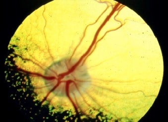
Optic neuritis is inflammation of the optic nerve. The optic nerve is the nerve that carries sight information from the eye to the brain (much like a USB cable takes information from a keyboard to a computer). Optic neuritis can mean the eye is working normally, as is the brain, but communication between the two is disrupted at some level. If the optic neuritis is not apparent in the eye then further tests are necessary (e.g ERG or MRI scans).
6. Brain blindness
This can happen without warning, or can happen after another brain injury (e.g epilepsy, trauma or anaesthesia). Often in these cases eye exams are normal. A diagnosis likely needs specialist images (MRI) and involvement of a veterinary neurologist. Treatment(s) depends on cause(s).
What diagnostic tests are used when trying to determine the cause of sudden onset blindness?
Electroretinography (ERG)
This measures the electrical activity of the light sensitive cells in the retina. A mild sedation is used to keep the animal still enough to perform the test. The response of the retinal cells produces a waveform which the ophthalmologist can interpret.
Ultrasound
An ultrasound scan using a specific probe designed for ocular use is a useful tool in diagnosing diseases such as cataracts and retinal detachment or for trauma cases. Some can be done under local topical anaesthesia, however usually a mild sedation is required so a more detailed ultrasound can be performed without stressing the patient.
Red/Blue lights
The retina and pupil respond to different colours of light in different ways. The ophthalmologist may shine a variety of coloured lights into the patient’s eyes to help with their diagnosis.
Maze Test
This involves allowing the patient to navigate an obstacle course in both light and dark conditions. The obstacles are frequently moved in different positions throughout the course so that the patient does not ‘learn’ the easy route. Specialised dog goggles (doggles) are used to cover one eye at a time before the maze test is conducted again, to allow the ophthalmologist to determine if vision is better in one eye over the other.
MRI/CT scans
Sometimes a more detailed scan of the patient is needed, for example in cases where ophthalmic examination results are normal. MRI and CT scans can be used to determine if there are any central nervous system, or brain issues which are causing the blindness (e.g optic neuritis or tumours). These scans are incredibly detailed and allow a 3-dimensional (in some cases 4-dimensional!) view of the area being scanned. Specialised radiographers will take these scans and interpret them. Your pet will be referred to a specialist centre to have these tests performed.
