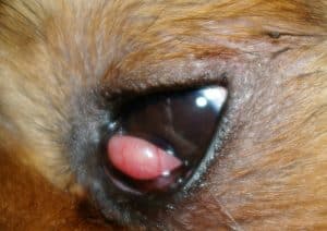
Cherry Eye
What is ‘Cherry eye’?
In addition to the upper and lower eyelids dogs and cats have a third eyelid on each eye. This has a protective function – taking the brunt of any trauma and hopefully sparing the eye itself from damage – and is also responsible for spreading the tear film across the eye surface. We call this eyelid the third eyelid or nictitating membrane.
The third eyelid has a cartilaginous frame and a gland at its base, which is responsible for producing 30-50% of the aqueous tear film. The gland is very loosely attached to the third eyelid and in some dogs it pops up from behind the edge of the third eyelid, looking like a red cherry hence its nickname ‘Cherry eye’. Although this looks dramatic it is not painful, but the longer it is exposed in this position the more irritated the gland and eyelid becomes, causing conjunctivitis and increased ocular discharge. The resulting irritation may cause the dog to rub at the eye and damage it, resulting in bleeding or infection. It may also cause reduced tear production.
‘Cherry eye’ is a condition usually seen in young dogs, typically when they are less than a year old. The breeds most commonly affected are Bulldogs, Cocker Spaniels, Shih Tzu, Lhasa Apsos, Poodles and Beagles. It is occasionally seen in Burmese cats. It usually affects both eyes but often not at the same time-the second gland may prolapse several months after the first.
Treatment
Surgical repair is required to correct this defect, although the ophthalmologist may prescribe topical anti-inflammatory medication for a period before this to reduce the inflammation first. In the past, the gland was actually removed – in a very quick procedure- which unfortunately tends to result in dry eye later in life. This requires life long medical treatment, and even further surgery to try to keep the eye lubricated. We now only advise removal if we are concerned about cancer in this area.
The technique most commonly used to repair this condition is the mucosal pocket technique. This requires a general anaesthetic and we make a small pocket on the inner aspect of the third eyelid, where the gland relocates, and then we stitch the area shut with dissolving sutures. The orbital rim technique may be used where permanent sutures are used to stitch the gland to the bone around the eye.
Unfortunately, no method is 100% effective and 10% of cases need repeat surgery. Re-prolapse is most common in animals which have had previous surgery in the area. Post-operative complications include infection, haemorrhage, re-prolapse, suture irritation of the cornea and cyst formation. Post-surgical inflammation may take 1-2 weeks to resolve.
Scrolling of the cartilage of the third eyelid
This condition has a very similar appearance to “cherry eye” and is often seen with a prolapsed gland. The cartilage frame inside the third eyelid is in a T shape, with a broad vertical band leading up to a thin horizontal portion. In some breeds, the broad vertical section kinks and this folds the third eyelid. The most commonly affected breeds are large dogs such as Great Danes, Weimeraners, Saint Bernards and Newfoundlands.
