Cataracts
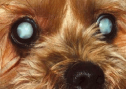
All animal eyes have a lens inside them. This lens helps the eye focus. It is a large structure shaped like two saucers placed together which is in the centre of the eye. Unlike the hard glass lens of a camera it is more like a clear jelly in a bag of cling film. A cataract is when this jelly goes cloudy or white.
What Causes Cataracts?
There are many causes of cataracts. These include; injuries to the eye, inflammation in the eye and diseases such as diabetes. There are also inherited types of cataract. Cataracts can also form due to diseases of other parts of the eye such as the retina.
What is the treatment for Cataracts?
There is no medical treatment known to slow the progression, prevent the formation or reverse the changes of cataracts. Like the egg , once “cooked” the white stays white. Surgery to remove the affected lens is the only treatment to provide a return to vision.
Should my pet have cataract surgery?
Cataract surgery is generally advised for those animals who have become blind because they have developed cataracts in both eyes. Not all animals or their eyes are suitable for surgery. In view of this an important part of the process is the Cataract Assessment. This is done under sedation.
Here the eyes are examined to make sure there is no other disease process present which might affect the outcome of the surgery. The retina (the light sensitive layer at the back of the eye) is examined using an ultrasound probe. This enables us to see the shape and size of the lens.
It is important to know whether the retina is working properly because we don’t want to end up in a situation that the animal cannot see once the cataract has been removed because the retina was already damaged. An electroretinography (ERG) will determine the retinal function of the eye and tell us if the retina is working or not.
A high percentage of cataracts tend to be in the older pet. It is therefore useful to know how fit an animal is prior to lengthy surgery. An assessment of this fitness can be done with tests on a blood sample. These tests are also useful if diabetic cataracts are suspected.
What does the surgery involve?
The dog’s eye reacts violently to any surgery. The dog’s lens is much bigger than a human lens and to remove it surgically would require a large hole. We use a technique called phacoemulsification. Here a special probe is passed into the eye. This vibrates, breaks up the cataract and then vacuums it out. This method causes least disturbance to the eye and hopefully the least problems. Unlike the human eye, when a dog is anaesthetised the cornea (clear part) disappears from view. To overcome this and allow the surgery a special anaesthetic is used. This temporally paralyses the eye muscles. However it also paralyses the breathing muscles so the anaesthetist has to connect the animal to a machine to breathe for it throughout the operation.
We like your pet to return home the same day if possible as they are usually more relaxed there. Post operative care involves frequent applications of multiple eye drops and oral tablets. Each of these eye drops will need to be given anywhere between 4 and 6 times daily. Your pet will be provided with a buster collar to wear for 2 weeks. This is to prevent any trauma to the eyes and is imperative to the success of the surgery.
What is the success rate?
With the advances of techniques, specialised equipment and drugs the success rate in dogs is 75-85% . Ten years ago it was 50-60%. Fortunately these techniques mean there are very few complications during the actual surgery.
Most of the complications occur in the days after surgery. These include bleeding into the eye. This particularly occurs if the animal gets excited and raises its blood pressure. The problems arise with major bleeding , this may take weeks to resolve and as it clots may pull off the retina.
The eye may develop glaucoma – a high pressure within the eye. If detected this needs be treated before it does damage to the eye. Further treatments or procedures may be needed to help control the intraocular pressures and you may need to bring your pet back for check-ups more regularly during this period.
The retina is held on the walls of the eye mainly by the fluids inside. Removing a large volume lens can allow the retina to loosen and detach. Thus post operative checks are essential to recognise and treat complications as they occur. This is usually in the form of eye drops or tablets but occasionally the animal has to be taken back to surgery.
What can your pet see after surgery?
Removal of the lens means that animals have a fixed focus at about 10 feet. As animals do not have the need for fine focus such as reading this sight is usually adequate for a happy life. Synthetic lenses may be fitted in dogs to improve focus. These are now available and can be implanted if the eyes are suitable.
In the immediate post-operative period there is a lot of inflammation within the eye which needs to be controlled. This is where the eye drops are important to reduce this inflammation and clear the visual axis for your pet. Any missed or forgotten drops can be detrimental to your pet’s surgical success.
