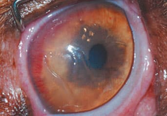
SCCED
What is a SCCED?
SCCED stands for spontaneous chronic corneal epithelial defect.
These are superficial corneal defects (“erosions”) which fail to heal spontaneously. The cornea is the transparent window at the front of the eye. It is a very thin area of tissue only 0.6mm thick which is made up of 3 different layers.
The disease occurs when the outermost corneal layer(epithelium) fails to adhere to the underlying middle layer (stroma).
What are the symptoms of a SCCED?
The corneal epithelium is highly packed with nerves and for this reason, patients with SCCEDs are often very painful. Increased blink rate (blepharospasm), squinting, increased lacrimation (tear
production) and a cloudy colour to the cornea are common signs.
Ulcers which fail to heal with medical treatment alone and become ‘static’ are commonly seen.
What is the cause?
The underlying cause is not completely clear. In a healthy cornea, an uncomplicated superficial ulcer heals usually in 5-7 days. In corneas where a SCCED is diagnosed, the epithelium grows over the defect but does not adhere to the underlying layer.
This causes a persistent corneal lesion which appears to ‘never heal’.
How can it be treated?
The goal of the treatment is to remove all the nonadherent epithelium and to stimulate and promote the adhesion of new epithelium.
Topical anaesthetic drops are used to anaesthetise the cornea and a sterile cotton bud is used to peel off the non-adherent epithelium. A diamond burr is then used to clean the bed of the defect. This treatment can be done either consciously or under sedation depending on the cooperation of the patient.
A contact lens is placed over the debrided cornea to provide comfort. Where animals have been sedated, a temporary tarsorrhaphy (1-2 stitches in the cornea of the eyelids) is performed to help the contact lens to stay in place.
Following debridement with a diamond burr, 92% of SCCEDs heal in 2 weeks, however a repeat second debridement may be necessary. Rarely (about 2% of cases) the diamond burr debridement is unsuccessful and a full superficial keratectomy under general anaesthetic is needed.
What happens on the day of the examination / surgery?
Your pet will be booked in for the procedure under sedation should a conscious approach not be appropriate. Your pet will be admitted to the clinic as a day patient and will be discharged later that day.
All pets receiving a sedation or anaesthetic are required to be starved overnight (unless younger than 3 months old). If your pet is diabetic, please speak to the ophthalmologist or nurse regarding your pet’s feeding/insulin regime prior to a sedation or anaesthetic.
What to do at home?
A buster collar needs to be worn for 2 weeks following the procedure. This is to prevent self-trauma by your pet rubbing at his/her eye as it heals. You will be provided with eye drops (antibiotics, lubrication)
along with tablets and pain relief for your pet.
It is imperative that you follow the written instructions regarding application of the drops and provision of the tablets to ensure your pet has the best chance of recovery. It is possible that the contact lens may fall out or be lost during the recovery period. Please contact us if this happens and we will advise you accordingly.
A check-up appointment is required in roughly 1 week to check the eye is healing as expected, and a further appointment 2 weeks after the initial treatment will be required to remove the contact lens and any sutures present.
