Lens Luxation
The lens is a clear structure located behind the iris inside the eye. It is held in place by lots of tiny tension cables called zonules. It helps to focus light onto the retina, where the light is transformed into an electrical signal and sent to the brain.
A lens luxation is a dislocation of the lens. In some breeds of dogs, usually Jack Russell Terriers and other terrier breeds, there is an underlying genetic defect causing the zonules to snap. This is called primary lens luxation.
Secondary lens luxation occurs as a consequence of other eye diseases, where there is inflammation inside the eye causing degeneration to the lens zonules. It can also happen if a disease causes the eye to increase in size, tearing the zonules as the eye stretches.
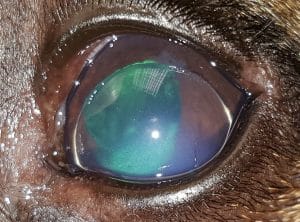
We also classify the luxation depending on which direction the lens falls. If the lens falls forward into the anterior chamber and sits in front of the iris we call it an anterior luxation. This causes acute pain because the lens suddenly blocks the drainage route of the fluid inside the eye, causing raised intra-ocular pressure (glaucoma). It is this increase in pressure which permanently damages the retina and causes blindness. The pressure rise also makes the front of the eye (the cornea) cloudy, as the damage to the corneal endothelium stops the tiny drainage pumps from moving water out of the cornea.
If the lens falls backwards and sits behind the iris we call it a posterior luxation. This tends not to cause an acute problem but can allow the gel in the back of the eye (the vitreous) to move through the pupil and block the drainage route. The lens damages the retina and can cause it to detach from the eye wall.
Lens luxation is a painful disease which can cause blindness if not diagnosed and treated quickly and effectively.
Clinical signs
These animals usually develop a very painful red and cloudy eye rapidly. This is often misdiagnosed as a conjunctivitis which fails to respond to treatment. The white part of the eye (sclera) becomes red and the clear window at the front of the eye (cornea) becomes cloudy/blue. These dogs show their pain by keeping the eye shut and becoming lethargic and depressed.
Your vet can see the lens sitting in the front of the iris with their ophthalmoscope. If the lens falls backwards, they can see a gap between the lens and the iris. As the pressure rises however, it becomes more difficult to see inside the eye, and more specialist equipment is needed to make the correct diagnosis. An ophthalmologist can use this equipment to spot the very subtle early signs of lens luxation before the lens falls out of position.
Treatment
If diagnosed early enough, medical therapy can be used to stop the lens falling through the pupil. Once the lens has dislocated, surgical therapy is required to correct this condition. Under general anaesthesia, the lens can be moved behind the iris, before topical drops are used to close the pupil and keep the lens in place. Alternatively the lens can be surgically removed through a small opening in the cornea, using an operating microscope and microsurgical instruments. If the luxation is not treated early enough, and blindness results because of glaucoma, it is sometimes necessary to remove the eye.
It is important to medically treat the unaffected eye to prevent luxation, as this disease is often bilateral (affects both eyes).
