Corneal Cross-Linking (CXL)
Focus on corneal collagen cross-linking (for referring vets).
What is corneal cross-linking (CXL)?
The normal cornea is comprised of collagen fibres and has enhanced biomechanical stability due to the presence of natural covalent cross-links between these fibres. In certain corneal diseases where the stability of the cornea is compromised, additional cross-links can be introduced using UV-A/Riboflavin corneal cross-linking. The procedure also has an antimicrobial effect and increases the resistance of collagen fibres to enzymatic degradation.
What do we use it for?
In veterinary ophthalmology the most common use for Corneal CXL is in the treatment of keratomalacia (melting ulcers). It may be used as the primary treatment modality along with medical management, or it may be used to stabilise the cornea prior to surgical grafting in extensive or deep ulcers.

CXL technique
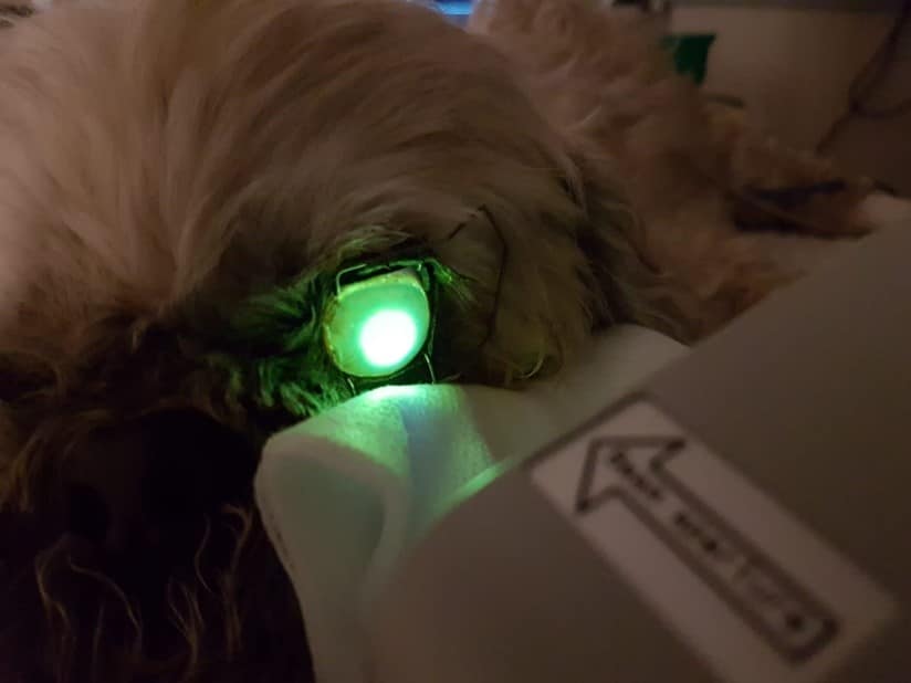
This technique requires specialist equipment which costs in excess of £10,000 and is used by our referral ophthalmology clinicians. CXL is usually performed in the sedated or anaesthetised animal however if for any reason this is contra-indicated, it can be performed conscious in an amenable patient. Dorsal or sternal recumbency is preferred in the anaesthetised patient and in either case the eyelids are held open using an appropriate speculum. The corneal surface is primed with Riboflavin which is applied at a rate of one drop every 2 minutes for a 30 minute period. The cornea is then flushed with sterile saline or balanced salt solution and anaesthetised topically. The cornea is then irradiated using UV-A light so that a total irradiance of 90 mW/cm2 is achieved.
Reference
Pot SA, Gallhofer NS, Metheis FL et al. Corneal collagen cross-linking as a treatment for infectious and non-infectious corneal melting in cats and dogs: results of a prospective, non-randomised, controlled trial. Veterinary Ophthalmology 2015; 17, 4: 250 – 260.